Wikisage is op 1 na de grootste internet-encyclopedie in het Nederlands. Iedereen kan de hier verzamelde kennis gratis gebruiken, zonder storende advertenties. De Koninklijke Bibliotheek van Nederland heeft Wikisage in 2018 aangemerkt als digitaal erfgoed.
- Wilt u meehelpen om Wikisage te laten groeien? Maak dan een account aan. U bent van harte welkom. Zie: Portaal:Gebruikers.
- Bent u blij met Wikisage, of wilt u juist meer? Dan stellen we een bescheiden donatie om de kosten te bestrijden zeer op prijs. Zie: Portaal:Donaties.

Bestand:614px-Eye-diagram svg.png: verschil tussen versies
({{Afbeelding |Beschrijving=Diagram van een oog : <br> 1:achterste oogkamer 2: ora serrata 3: musculus ciliaris 4: zonula ciliaris 5: kanaal van Schlemm 6: pupil 7: [[voorste oogkam) |
Geen bewerkingssamenvatting |
||
| Regel 1: | Regel 1: | ||
{{Afbeelding | {{Afbeelding | ||
|Beschrijving=Diagram van een [[oog (anatomie)|oog]] : <br> 1:[[achterste oogkamer]] 2: [[ora serrata retinae|ora serrata]] 3: [[musculus ciliaris]] 4: [[zonula ciliaris]] 5: [[kanaal van Schlemm]] 6: [[Pupil (oog)|pupil]] 7: [[voorste oogkamer]] 8: [[hoornvlies]] 9: [[Iris (anatomie)|iris]] 10: [[Ooglens|lens kapsel]] 11: [[Ooglens|lenskern]] 12: [[processus ciliares]] 13: [[conjunctiva]] 14: [[musculus obliquus inferior|m. obliquus inferior]] 15: [[musculus rectus inferior]] 16: [[musculus rectus medialis]] 17: [[retinale arteriën en venen]] 18: [[blinde vlek]] 19: [[harde hersenvlies]] 20: [[arteria centralis retinae]] 21: [[vena centralis retinae]] 22: [[nervus opticus]] 23: [[venae vorticosae]] 24: [[harde oogrok]] 25: [[gele vlek]] 26: [[fovea centralis]] 27: [[sclera]] 28: [[vaatvlies]] 29: [[musculus obliquus superior|m. obliquus superior]] 30: [[netvlies]] | |Beschrijving= Diagram van een [[oog (anatomie)|oog]] : <br> 1:[[achterste oogkamer]] 2: [[ora serrata retinae|ora serrata]] 3: [[musculus ciliaris]] 4: [[zonula ciliaris]] 5: [[kanaal van Schlemm]] 6: [[Pupil (oog)|pupil]] 7: [[voorste oogkamer]] 8: [[hoornvlies]] 9: [[Iris (anatomie)|iris]] 10: [[Ooglens|lens kapsel]] 11: [[Ooglens|lenskern]] 12: [[processus ciliares]] 13: [[conjunctiva]] 14: [[musculus obliquus inferior|m. obliquus inferior]] 15: [[musculus rectus inferior]] 16: [[musculus rectus medialis]] 17: [[retinale arteriën en venen]] 18: [[blinde vlek]] 19: [[harde hersenvlies]] 20: [[arteria centralis retinae]] 21: [[vena centralis retinae]] 22: [[nervus opticus]] 23: [[venae vorticosae]] 24: [[harde oogrok]] 25: [[gele vlek]] 26: [[fovea centralis]] 27: [[sclera]] 28: [[vaatvlies]] 29: [[musculus obliquus superior|m. obliquus superior]] 30: [[netvlies]] | ||
|Bron=[http://commons.wikimedia.org/w/index.php?title=File:Eye-diagram.svg&oldid=26577371 Wikimedia Commons] | |Bron=[http://commons.wikimedia.org/w/index.php?title=File:Eye-diagram.svg&oldid=26577371 Wikimedia Commons] | ||
|Datum afbeelding=3 Maart 2007 | |Datum afbeelding=3 Maart 2007 | ||
| Regel 11: | Regel 11: | ||
|Licentie=cc-sa | |Licentie=cc-sa | ||
}} | }} | ||
[[Categorie:Wikisage:Afbeeldingen:Ogen]] | |||
Huidige versie van 10 sep 2009 om 12:21
| Beschrijving: | Diagram van een oog : 1:achterste oogkamer 2: ora serrata 3: musculus ciliaris 4: zonula ciliaris 5: kanaal van Schlemm 6: pupil 7: voorste oogkamer 8: hoornvlies 9: iris 10: lens kapsel 11: lenskern 12: processus ciliares 13: conjunctiva 14: m. obliquus inferior 15: musculus rectus inferior 16: musculus rectus medialis 17: retinale arteriën en venen 18: blinde vlek 19: harde hersenvlies 20: arteria centralis retinae 21: vena centralis retinae 22: nervus opticus 23: venae vorticosae 24: harde oogrok 25: gele vlek 26: fovea centralis 27: sclera 28: vaatvlies 29: m. obliquus superior 30: netvlies |
| Bron: | Wikimedia Commons |
| Afbeelding gemaakt op/in: | 3 Maart 2007 |
| Afbeelding gepubliceerd op/in: | 3 Maart 2007 |
| Auteur: | Chabacano |
| Originele uploader: | Chabacano |
| Toestemming: | ja |
| Andere versies: | 200px, 500px, 1000px, 2000px, Cijfers niet omcirkeld, Rode achtergrond |
Licentie:
Dit materiaal is door de auteur onder de licentie Creative Commons - Share Alike, versie 1.0 gepubliceerd.
Dit materiaal mag vrij verspreid, gekopieërd en/of bewerkt worden op voorwaarde dat het materiaal en afgeleide materiaal onder deze licentie wordt gepubliceerd.Meer informatie
![]() This media has been published by Creative Commons - Share Alike, version 1.0 by the author.
This media has been published by Creative Commons - Share Alike, version 1.0 by the author.
This media may be published, copied and/or edited on condition that this media and diverted media will be published on this license. information
Bestandsgeschiedenis
Klik op een datum/tijd om het bestand te zien zoals het destijds was.
| Datum/tijd | Miniatuur | Afmetingen | Gebruiker | Opmerking | |
|---|---|---|---|---|---|
| huidige versie | 10 sep 2009 12:19 | 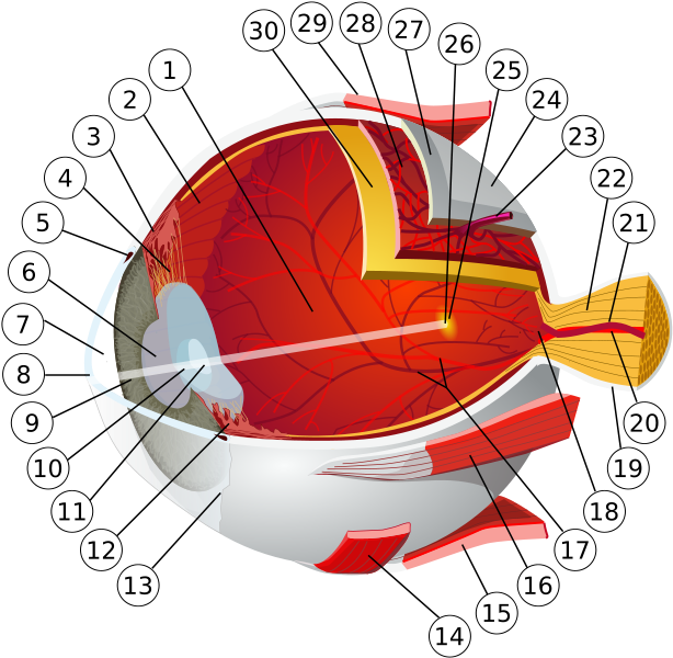 | 614 × 600 (254 kB) | SjorsXY (overleg | bijdragen) | {{Afbeelding |Beschrijving=Diagram van een oog : <br> 1:achterste oogkamer 2: ora serrata 3: musculus ciliaris 4: zonula ciliaris 5: kanaal van Schlemm 6: pupil 7: [[voorste oogkam |
U kunt dit bestand niet overschrijven.
Bestandsgebruik
Dit bestand wordt op de volgende pagina gebruikt: